
Services

CT Scan of the Chest

A chest CT gives your physician detailed images of the lungs, airways, blood vessels, and other structures in your chest. It can help investigate respiratory symptoms, evaluate abnormalities, and provide a clearer picture of conditions affecting the chest.

What Is a CT Scan of the Chest?

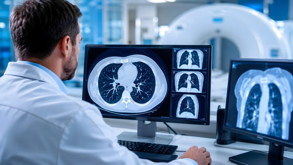
A CT scan of the chest is a non-invasive imaging exam that uses X-rays and computer technology to create detailed images of the structures inside the chest. Unlike a standard X-ray, CT captures multiple views that can be reconstructed into cross-sectional images of the lungs and surrounding anatomy.

Your physician may recommend a chest CT when a standard chest X-ray does not provide enough detail or when a known or suspected condition needs further evaluation. Depending on the reason for the exam, the scan may be performed with or without IV contrast.

Your physician may order a chest CT to investigate persistent respiratory symptoms, evaluate an abnormality seen on another imaging exam, monitor a known condition, or assess the chest following an injury.

Why Choose Gateway Diagnostic Imaging for Your Chest CT?

Gateway Diagnostic Imaging provides affordable, high-quality CT imaging at convenient outpatient locations throughout the Dallas-Fort Worth metroplex and Abilene. We’re in-network with all major insurance plans, and patients can save up to 93% compared to hospital imaging costs without compromising quality care or diagnostic accuracy.

With multiple locations and same-day or next-day appointments often available, Gateway makes it easy to schedule your chest CT when you need it. Your images are reviewed by board-certified, subspecialized radiologists, and the results are sent to your referring physician.

What Does a CT Scan Show in the Chest?

A chest CT can provide detailed views of many structures within the thoracic cavity, including the:

  • Lungs
  • Bronchi and other airways
  • Aorta and other major blood vessels
  • Mediastinum
  • Lymph nodes
  • Pleura
  • Ribs and other portions of the chest wall
  • Upper portions of the abdomen visible on the scan

Your physician may use chest CT imaging to evaluate:

  • Persistent cough or unexplained respiratory symptoms
  • Shortness of breath
  • Lung nodules or other pulmonary abnormalities
  • Pneumonia and other infections
  • Inflammation or scarring within the lungs
  • Emphysema and other chronic lung changes
  • Enlarged lymph nodes
  • Abnormal findings identified on a chest X-ray
  • Chest injuries or trauma
  • Suspected tumors or other masses
  • Changes associated with a known medical condition

The information from the scan can help your physician distinguish between different possible causes of symptoms and determine whether further evaluation or treatment is needed.

How To Prepare for a CT Scan of Your Chest

Scheduling Considerations

  • Same-day and next-day appointments may be available.
  • Preparation depends on whether your exam requires contrast.
  • Follow any fasting or medication instructions provided before your appointment.
  • Arrive approximately 15 minutes before your scheduled exam.

Medical Information to Provide

  • Inform the imaging team about allergies or previous reactions to contrast material.
  • Provide a current list of medications and relevant medical history.

Insurance and Documentation

  • Bring your insurance card.
  • Bring a valid photo ID.

What to Wear

  • Wear comfortable clothing that does not contain unnecessary metal.
  • You may be asked to remove jewelry, glasses, watches, or other removable items.
  • Depending on the exam, you may be provided with a gown.

What To Expect During the CT Scan of Your Chest

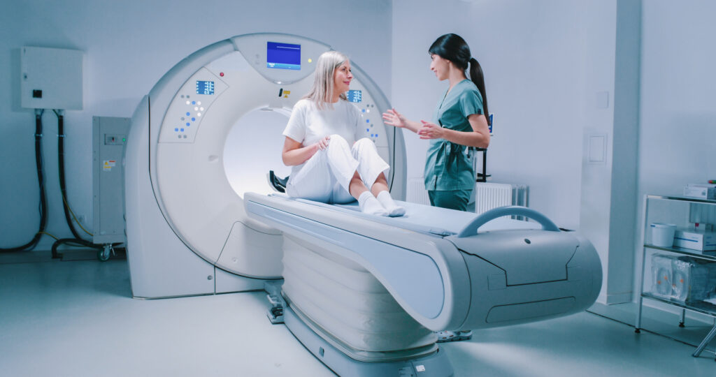
After checking in, a technologist will go over the plan and position you on the CT table. You will typically lie on your back while the table moves through the scanner.

During image acquisition, you may be asked to hold your breath briefly. Remaining still and following these breathing instructions helps produce sharper images of the lungs and other structures.

If IV contrast is part of your exam, the technologist will place an IV and administer the contrast during the scan. You may briefly feel warm or notice an unusual taste when the contrast is injected.

The imaging portion of a chest CT is usually completed within several minutes. Your appointment may take longer if contrast or additional preparation is required.

What To Expect After Your Chest CT Scan

Most patients can leave the imaging center and return to their normal routine immediately after a chest CT. If you received contrast, the imaging team may recommend drinking fluids afterward and will provide any additional instructions that apply to your examination.

Your images are evaluated by a board-certified, subspecialized radiologist. The radiologist prepares a report describing the imaging findings, which is sent to your referring physician. Your physician will review the results with you and determine what they mean in the context of your symptoms, medical history, and other test results.

FAQs

Where are Gateway Diagnostic Imaging chest CT scans available?

Gateway Diagnostic Imaging offers outpatient CT imaging at centers throughout the Dallas-Fort Worth metroplex and Abilene, providing convenient access to diagnostic imaging without a hospital-based setting.

How long does a chest CT scan take?

The imaging portion of a chest CT typically takes only a few minutes. Your total appointment time may be longer if contrast is required or additional preparation is needed.

Is a chest CT used for lung cancer screening?

Yes. CT can be used to screen for lung cancer in people who meet certain risk and eligibility criteria. Gateway Diagnostic Imaging offers specific, low-dose CT lung cancer screening as a separate service. 

Ready to Get Started?

Schedule a Chest CT scan today.

Find A Location Near You

Find A Location

Request An Appointment

Appointment Form